Press Releases
May 18, 2016
JAMSTEC
Symbiotic Bacteria Found on Outer Surface of Deep-sea Clam Eggs
-Successful on-board spawning induction of Calyptogena Okutanii -
Overview
A research group led by Dr. Tetsuro Ikuta at Department of Marine Biodiversity Research, the Japan Agency for Marine-Earth Science and Technology (JAMSTEC: Asahiko Taira, President) succeeded in inducing spawning of the deep-sea clam, Calyptogena Okutanii (C. okutanii : figure 1) in the laboratory. The result revealed extremely unique localization of the symbiont associating with the eggs.
Deep-sea giant clams of the genus Calyptogena live in hydrothermal vent or seep sites. Harbouring obligate sulfur-oxidizing intracellular symbiotic bacteria in the gill, this mysterious deep-sea clam obtains all the necessary nutrition from the symbiont. Calyptogena clams have been believed to transmit symbiotic bacteria via eggs from generation to generation. However, because of difficulty in sampling of the eggs, the question how the symbiont associates with the spawned eggs has remained unanswered.
To understand the processes underlying symbiotic associations and their evolution, the research group has established a method for artificial on-board induction of spawning of the Calyptogena okutanii, and investigated localization of the symbiont associating with spawned eggs, using the clams collected during JAMSTEC's cruise by ROV Hyper Dolphin at a depth of about 1,000 m in the Mid-Okinawa Trough and off Hatsushima in Sagami Bay. As a result, they found that injection of serotonin into the foot of the clam was effective for spawning. It is the first successful experiment of artificial spawning of Calyptogena clams in a laboratory.
Moreover, they revealed that 1) the symbiotic bacteria of C. okutanii are located at the egg's vegetal pole, being attached extracellularly on the outer surface of the egg plasma membrane and 2) an individual egg carries on average about 400 symbiont cells, each of which contains close to 10 genomic copies. Such extracellular localization of the symbiont on the egg should represent an unprecedented novel mode of the transmission of 'intracellular' symbiont. These findings shed light on the mechanisms of symbiont transmission and the genome evolution of symbionts, and also bring a new dimension to the embryological study of chemosymbiotic animals.
The above results were published on Royal Society Open Science on May 18. 2016 (JST).
Title: Surfing the vegetal pole in a small population: extracellular vertical transmission of an 'intracellular' deep-sea clam symbiont
Authors: Tetsuro Ikuta1, Kanae Igawa1,2, Akihiro Tame3, Tsuneyoshi Kuroiwa4,5, Haruko Kuroiwa4,5, Yui Aoki1, Yoshihiro Takaki6, Yukiko Nagai1, Genki Ozawa1,6, Masahiro Yamamoto1, Ryusaku Deguchi7, Katsunori Fujikura1, Tadashi Maruyama1,
Takao Yoshida1,2,6
Affiliations:
1 JAMSTEC 2 Tokyo University of Marine Science and Technology 3 Marine Works Japan, Ltd. 4 Japan Women's University 5 Japan Science and Technology Agency 6 Kitasato University 7 Miyagi University of Education
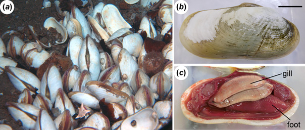
Figure 1. C.okutanii harbouring obligate symbiotic bacteria in the gill epithelial cells.
(a) A colony of C.okutanii in deep-sediments (at a depth of 1,055m in the Mid-Okinawa Trough (b) External appearance of the clam in the scale of 2cm. (c) Inside of the shell. A reddish-white and long structure is the gill, where symbiotic bacteria colonize and provide all the necessary nutrition to the host. The gonad is located in the dorsal side of the foot between the right and left gills.
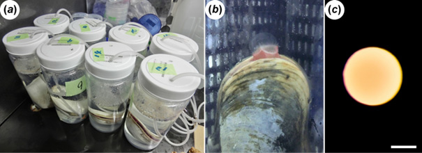
Figure 2. a) A snapshot of on-board spawning induction. Immediately after injection of serotonin in the foot, each clam was placed in a plastic container filled with 2 liter of seawater at 4°C with aeration. Spawning was observed after several ten minutes in only female clams with mature eggs. (b) A clam releasing eggs in a container. (c) A spawned egg with diameter of about 0.2 mm. Bar, 0.1mm.

Figure 3. (a) Localization of the C. okutanii symbiont associating with spawned eggs. The bluish-purple signal with small oval-shape shows a cluster of symbiotic bacteria (about 400 symbiont cells are there). Dark pink area indicates the vegetal hemisphere. (b-e) TEM (Transmission Electron Microscope) images of the symbiotic bacteria of C. okutanii on eggs. Animal side is up, and vegetal side is down. Red rectangles in b) and c) correspond to the locations of the following magnified images. (f) A schematic drawing showing localization of the symbiont on the egg surface. Red line indicates egg cell membrane. Yellow, dark blue and green areas indicate egg cytoplasm, symbiont and vitelline membrane, respectively. (g) Signals shown in light blue spots are symbiotic bacteria observed by a confocal microscopy. About 400 symbiont cells are observed in the oval-shaped area (100μm × 20 - 50μm). Bars in b), 50μm; c), 5μm; d), 0.5μm; e), 0.2μm; and g), 10μm.
Clamming in Deep Sea and On-board Spawning Induction
Contacts:
- JAMSTEC
- (For this study)
- Tetsuro Ikuta, Research Scientist, Department of Marine Biodiversity Research
- (For press release)
- Tsuyoshi Noguchi, Manager, Press Division, Public Relations