Imaging of nutrient uptake
It became into reality to demonstrate the subseafloor microorganisms activity in direct manner by using a nano-scale secondary ion mass spectrometer (NanoSIMS) as their incorporation of nutrients. Narrow (down to 50 nm) cesium ion beam hits micoorganisms and locally induce secondary ion generation. Then the generated secondary ions are separated based on their mass (weight of elements), and then the local elemental composition can be obtained and ion images are created by scanning the cesium ion beam across selected region. The carbon (12C) and nitrogen (14N) atom constituting the nutrients are replaced to stable isotopes (13C and 15N), so that its incorporation can be traced with NanoSIMS. By detecting increasing amount of stable isotopes, nutrient incorporation into microbial cell body can be imaged and quantitatively monitored at single cell-level. Since its starved and thus slow metabolic nature of subseafloor microorganisms, as well as their life in it had been very difficult to study subseafloor microorganisms with existing technologies. Dr. Morono and his colleagues also came up with another new technologies and adapted them to the subseafloor environment, made it capable to enumerate the microorganisms specifically and analyze their DNA quantitatively. With a capability to measure the metabolic activity by NanoSIMS, Dr. Morono is excited about the possibilities for the next drilling. He says that a technical capability is now in place that makes it possible to come into closer and closer for exploring subseafloor biosphere as a largest microbial habitat on Earth. Starting in the summer of 2012, the CHIKYU plans to drill again off the coast of Hachinohe and conduct detailed microbiological analyses onboard the vessel. There is a hope that the drilling will result in completely new data that can be the key in unlocking mysteries about the interaction between microorganisms and energy resources, and about the evolution of life.
 |
 |
|
Nano-scale secondary ion mass spectrometer (NanoSIMS) |
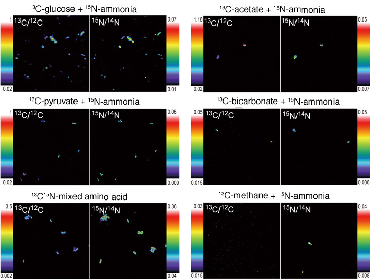
NanoSIMS images of microbial cells that have taken up nutrient resources labeled with stable isotopes of carbon (13C) and nitrogen (15N). NanoSIMS analysis makes it possible to make visible the elemental content at a spatial resolution as small as 50 nanometers (one nanometer = one-millionth of a millimeter). Given this capacity, it is possible to measure the elemental content of a single cell as small as 0.5–1 micrometer in size (1 micrometer = one one-thousandth of a millimeter). |
![]()
Kochi Institute for Core Sample Research
 |
 |
|

- |1|
- 2|